|
|
|
|
|
|
|
|
|
|
|
|
|
|
|
|
|
|
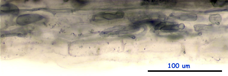
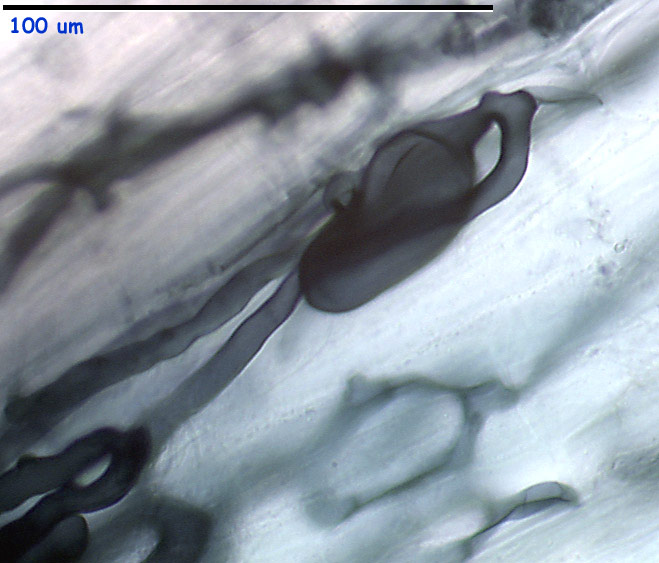
The first subtype is the classical, badly staining type where
mostly only the entry point hyphae and adjacent irregularly shaped (intracellular)
vesicles are visible. Rarely, the arbuscules are seen, but badly stained,
usually placed in a deeper layer of cortical cells.
The images are improved using the image-processing software, the original contrast of hyphae and vesicles was even smaller.
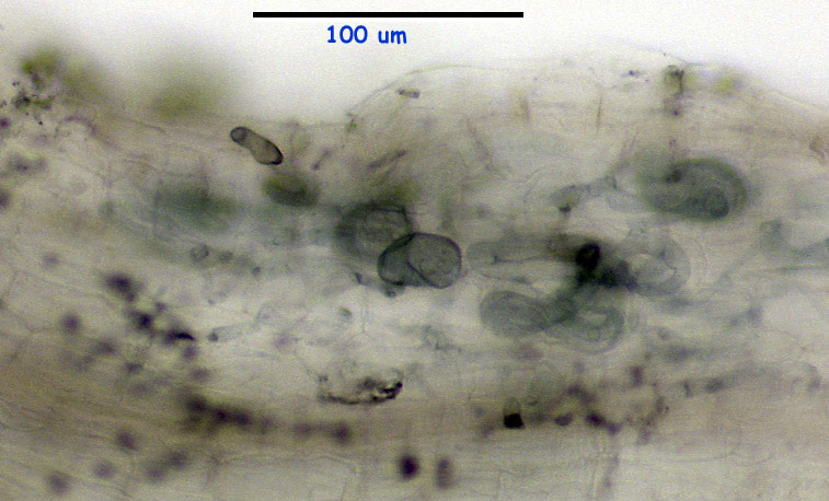
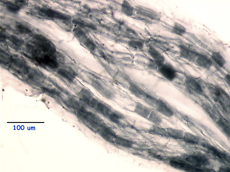
The second "Acaulospora" subtype seems to be quite similar
to the previous, but stains well, usually "too well". While I feel this
type is distinct from the plG3 ("T7") type, the exact divide line is very
difficult to draw between these two.
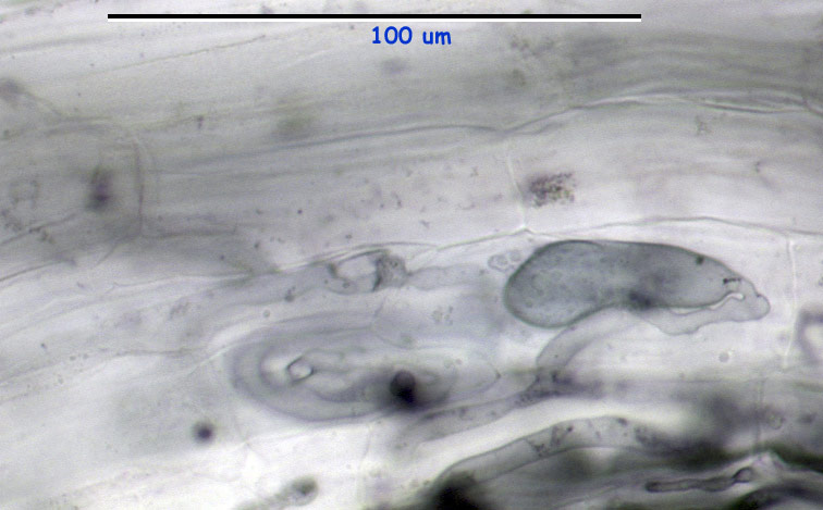
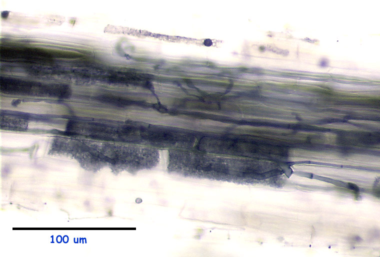
A vesicle detail:
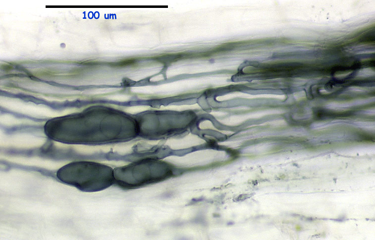
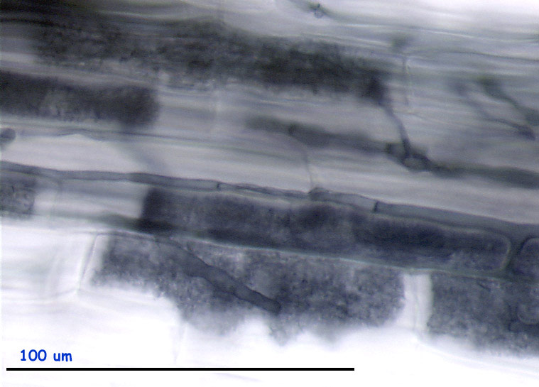
The last "Acaulospora" subtype is represented mainly
by the "brick-like" arbuscules, completely filling the cells. Sometimes
the parallel, longitudinally running hyphae are seen, but hardly any complex
hyphal structures or vesicles.
This page last modified by Petr milauer on 14 September 2000