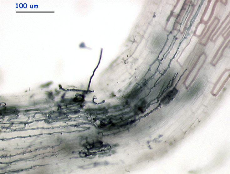
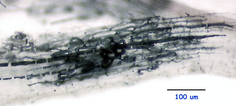
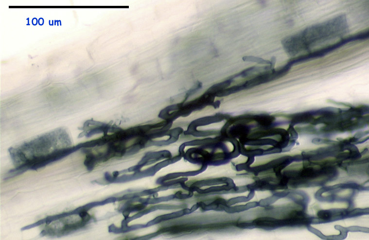
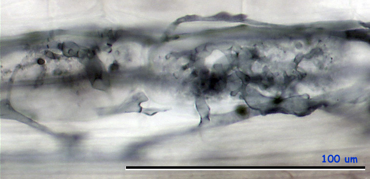
The images above and below represent a typical look of the starting Scutellospora colonies.
|
|
|
|
|
|
|
|
|
|
|
|
|
|
|
|
|
|
The images above and below represent a typical look of the starting
Scutellospora colonies.
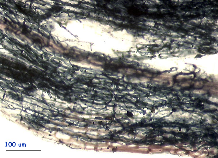
Following image represents a much developed Scutellospora colony (probably
originating from several entry points), with the hyphal coils filling all
the root segment volume, hidding details like arbuscules development or
eventual AM symbionts present in the deeper cortex layers.
A typical look of the plSc morphotype arbuscules:
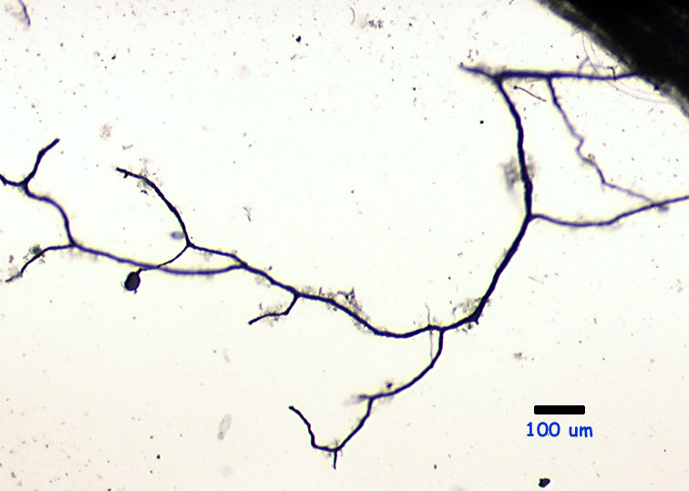
A branched extra-radical hypha emanating from a colony within Plantago
root and bearing one auxiliary cell (the other were probably un-plugged
during the sample processing)
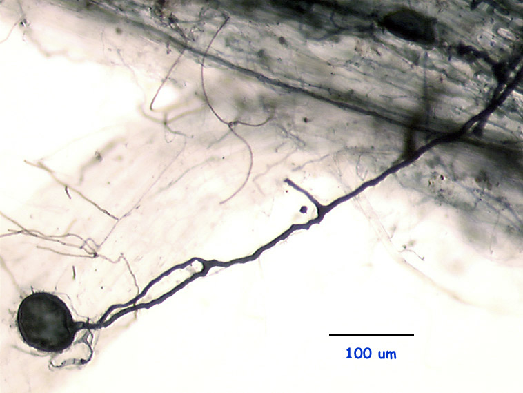
Detail of the auxiliary cell from another sample:
This page last modified by Petr milauer on 14 September 2000